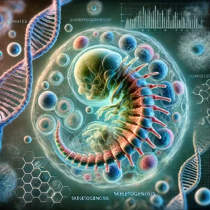
Somitogenesis and skeletogenesis are two essential biological processes that underlie the formation of the body’s axial skeleton and segmented musculature. These processes are responsible for the development of somites—segmented blocks of mesodermal tissue—during early vertebrate embryogenesis. While they are fundamental to shaping the vertebrate body plan, our understanding of the genetic and cellular mechanisms that govern somitogenesis and skeletogenesis has only started to come into focus in recent years. This blog explores the genetic pathways involved in somitogenesis, the transformation of mesodermal cells into somites, and the key genes that control vertebral and skeletal development.
Understanding Somitogenesis: A Complex Cellular Process
Somitogenesis is a critical process that occurs in the developing embryo, leading to the formation of somites from the presomitic mesoderm. Somites are precursors to various tissues, including the vertebrae, skeletal muscles, and dermis. The process of somitogenesis is highly regulated and involves a series of precise cellular and molecular events, including cell migration, differentiation, and changes in cell adhesion.
One of the fascinating aspects of somitogenesis is the transition that mesodermal cells undergo as they move from an epithelial to a mesenchymal state. During early somitogenesis, mesodermal cells exhibit significant changes in cell adhesion. Research has shown that somitic cells are more self-adherent than those in the presomitic mesoderm, suggesting that cell adhesion plays a vital role in the organization and segmentation of somites. This cell adhesion phenomenon is crucial for the correct positioning and patterning of somites along the anterior-posterior axis of the developing embryo.
The Mesodermal Transition: From Mesenchymal to Epithelial
During the formation of somites, mesodermal cells undergo a dramatic shift in their morphology and behavior. Initially, mesodermal cells are loosely arranged and mesenchymal in nature. As somitogenesis progresses, these cells transition into an epithelial state—characterized by tight cell-cell junctions and a more organized structure. This transition is essential for the formation of well-defined somites.
In the subsequent stage, known as sclerotome formation, cells revert to a mesenchymal state, which allows for the development of vertebral precursors. This mesenchymal-to-epithelial and epithelial-to-mesenchymal transition is a hallmark of vertebrate development, especially in the context of axial patterning and skeletal formation.
Somitogenesis in Vertebrate Models: Chick, Xenopus, Mouse, and Zebrafish
Over the past five decades, the study of somitogenesis has been a focal point in developmental biology, with researchers using a variety of vertebrate model organisms to better understand this process. Model organisms such as the chick, Xenopus (frogs), mice, and zebrafish have provided invaluable insights into the genetic and molecular regulation of somite formation.
In particular, the mouse model has been instrumental in understanding the cellular dynamics of somitogenesis. In mice, somitogenesis occurs along the anterior-posterior axis of the developing embryo, typically between embryonic days E8 and E12. During this period, somites develop from the lateral mesoderm and progressively organize into the segments that will later differentiate into vertebrae and musculature.
A key feature of somitogenesis is the differentiation of somites along two major axes: the anterior-posterior axis and the dorsal-ventral axis. As somites form, heterogeneity develops between their anterior and posterior regions, leading to the segmentation of the body. Additionally, interactions with surrounding structures such as the notochord and neural tube create asymmetries within the somites, contributing to their final structural organization.
The Role of Genes in Somitogenesis: Key Genetic Pathways
Understanding the genes involved in somitogenesis has been a major scientific breakthrough. Many of the genes that control somitogenesis in vertebrates were first identified in Drosophila, the fruit fly, through genetic screens. Researchers have since discovered homologous genes in vertebrates that perform similar functions in regulating body segmentation.
The genetic pathways that govern somitogenesis can be grouped into several families, each playing a critical role in somite formation and patterning. Some of the most important gene families involved in somitogenesis include:
- Fibroblast Growth Factor (FGF) Family: FGF signaling is crucial for mesodermal cell proliferation and differentiation during somitogenesis.
- Hedgehog Family: The Hedgehog signaling pathway is essential for patterning and the differentiation of somites into specific structures.
- Wnt Family: Wnt signaling regulates cell fate decisions and contributes to somite formation.
- TGF-β/BMP Family: Transforming growth factor beta (TGF-β) and bone morphogenetic proteins (BMPs) are involved in the regulation of mesodermal cell differentiation and tissue patterning.
- Homeobox (Hox) Gene Family: Hox genes play a key role in specifying the regional identity of somites along the anterior-posterior axis.
- Pax Family: Paired box (Pax) genes regulate somite patterning and are important for the proper development of vertebrae and skeletal muscles.
- bHLH Gene Family: Basic helix-loop-helix (bHLH) transcription factors are involved in the regulation of myogenesis within somites.
- Notch Signaling Pathway: Notch signaling regulates the segmentation clock, which controls the periodic formation of somites.
Additionally, a small number of genes involved in somitogenesis have been identified through positional cloning efforts. These include classical mutants such as Brachury and Fused, which are linked to defects in tail vertebrae and somite development. These findings highlight the complexity and precision of the genetic networks that regulate somitogenesis.
Positionally Cloned Mutations and Vertebral Defects
Positional cloning has provided deeper insights into the genetic defects that lead to abnormal somitogenesis. Mutations affecting the development of tail vertebrae, in particular, have been extensively studied. These mutations often result in visible defects in the vertebral column, particularly in the tail region, which is composed of more than half of the vertebrae in the body.
Among the many positional clones identified, mutations in genes like Brachury and Fused have been linked to defects in the somite patterning and vertebral formation. These mutations serve as critical models for understanding how genetic disruptions in somitogenesis can lead to skeletal abnormalities and other developmental disorders.
Conclusion: The Future of Somitogenesis Research
The genetic mechanisms underlying somitogenesis are still being explored, with many exciting discoveries yet to be made. As we continue to uncover the specific roles of various gene families and signaling pathways, we gain a deeper understanding of how the body develops and organizes its complex structures. Moreover, these insights hold promise for advancing our knowledge of developmental disorders and congenital diseases that affect the vertebral column and musculature.
For science students and budding developmental biologists, understanding the intricacies of somitogenesis is not only crucial for grasping the fundamentals of developmental biology but also for tackling future challenges in regenerative medicine, genetic diseases, and tissue engineering.


